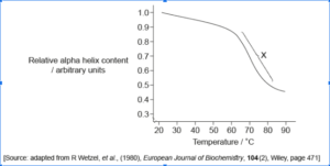
General Science IBDP Biology 7.3 Translation May 14, 2021 Shveta 0 Comments Biology, IBDP Biology, Nucleic Acids (AHL), Translation 1.Which cell component synthesizes actin and myosin?Free ribosomes Rough endoplasmic reticulum Smooth endoplasmic reticulum Nuclear membrane 2. Which structure found in eukaryotes has a single membrane?Nucleus Lysosome Chloroplast Mitochondrion3. Which types of interactions are found in a part of a protein with secondary but not tertiary structure? I. Hydrogen bonds II. Disulphide bridges III. Ionic bondsI only I and II only II and III only I, II and III 4. This image is taken from a visualization of a eukaryotic ribosome. The arrows show the direction of movement of mRNA. Which letter represents a tRNA exiting from the E site? ABCD5. Scientists have heated a solution containing the protein albumin and measured its relative alpha helix content, shown on the graph. What does the zone labelled X indicate?Rapid increase in beta pleated sheets Rapid formation of hydrogen bonds Rapid increase in denatured protein molecules Rapid decrease in peptide bonds6. Where does a tRNA-activating enzyme attach the appropriate amino acid to the tRNA molecule? ABCD7. What does post-transcriptional modification of eukaryotic mRNA include? What does post-transcriptional modification of eukaryotic mRNA include? I. Introns are removed from mRNA. II. Exons are joined together to form mature mRNA. III. A 5' cap and 3' poly-A tail are added to mRNA. I only . I and III only II and III only I, II and III 8. Variations in the types of antibodies are produced by mRNA splicing. What is an advantage of this process?Reduces the size of mRNA required for the translation of antibodiesIncreases the number of different antibodies that can be synthesized Ensures that one gene codes for one antibody Speeds up transcription of antibodies9. The image represents a model of the protein transthyretin. Which level of structure is indicated by X on the image?Primary Secondary Tertiary Quaternary 10. The image shows the structure of hemoglobin. What determines the primary structure of hemoglobin? Genetic information Hydrogen bonding Four polypeptide chainsSide chain interactions 11. The following diagram shows a ribosome during translation. What describes the specific stage of translation?Initiation Elongation Termination Translocation12.The diagram is a three-dimensional molecular model of a protein. Which bonds stabilize the shape of the area labelled X?Covalent bonds between adjacent amino acids Hydrogen bonds between N–H and C=O groups of amino acidsHydrophobic interactions between R groups of amino acids Disulphide bridges between cysteine molecules 13. The diagram shows the cross section of a plasma membrane. What is found in area X?Glycolipid GlycoproteinPolar amino acid Non-polar amino acid14. Which best describes the tertiary structure of a protein?The interaction of polypeptide subunits and prosthetic groupsInteractions forming hydrogen bonds between the amino acids The sequence of amino acids in the polypeptide chain The structure formed from interactions between the amino acid side groups 15. What is the distinction between highly repetitive DNA sequences and single-copy genes? The highly repetitive sequences have greater amounts of guanine The highly repetitive sequences have greater amounts of cytosineThe highly repetitive sequences are not transcribedThe highly repetitive sequences are not replicated