1.2- Ultrastructure of cells

1.The image is of a Paramecium   [Source: Adapted from www.biology-resources.com. Copyright 2004-2017 D G Mackean & Ian Mackean. All rights reserved.] Which function is accomplished by structures X and Y in the Paramecium?      


2. What evidence from the image of Paramecium indicates whether the organism is a prokaryote or a eukaryote?


3. [Source: Adapted from www.biology-resources.com. Copyright 2004–2017 D G Mackean & Ian Mackean. All rights reserved.] Which function is accomplished by structures X and Y in the Paramecium?


4. Which structure found in eukaryotes has a single membrane?


5. The following electron micrograph shows part of a palisade mesophyll cell. Which of the labelled structures controls the exchange of substances to and from the cell?


6. Cladograms can be created by comparing DNA or protein sequences. The cladogram on the left is based on DNA sequences and the cladogram on the right is based on comparing protein sequences.   What is the reason that cladograms based on DNA sequences are more reliable predictors of the phylogenetic relationship of species than cladograms based on protein sequences?


7. Which organism has DNA located in three organelles?


8. Which organelle is involved in generating vesicles destined for the cell membrane?


9. The image shows an electron micrograph of mesophyll cells. What is the name of the structure labelled X?


10. The bacterium Neisseria gonorrhoeae causes infections related to the human reproductive system. The graph shows the percentage of samples in which this bacterium showed resistance to six antibiotics over a period of ten years.     What is a possible explanation for the total percentage resistance being larger than 100% in 2010?


11. The image shows an electron micrograph of pancreatic exocrine cells.   What is the role of the vesicles shown in the micrograph?


12. What distinguishes prokaryotic cells and eukaryotic cells?


13. Which substance is used for structure in plants?


14. Animal cells often secrete glycoproteins as extracellular components. What is a role of these glycoproteins?


15. The image represents an Escherichia coli.     What is the function of structure X?


16. Animal cells often secrete glycoproteins as extracellular components. What is a role of these glycoproteins?


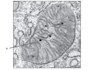
17. What is the structure labeled X in the electron micrograph of a rat liver cell?     [Source: “0315 Mitochondrion new” by OpenStax College - Anatomy & Physiology, Connexions Web site. http://cnx.org/content/col11496/1.6/, Jun 19, 2013.. Licensed under CC BY 3.0 via Wikimedia Commons - https://commons.wikimedia.org/wiki/File:0315_Mitochondrion_new.jpg#/media/File:0315_Mitochondrion_new.jpg]


18. What is a function of the plant cell wall?


19. What distinguishes prokaryotic cells from eukaryotic cells?


20. What structures are part of an Escherichia coli cell?


21. What is a function of the plant cell wall?


22. What distinguishes prokaryotic cells from eukaryotic cells?


23. What is an example of binary fission?


24. Where are proteins synthesized by free ribosomes used?


25. What are characteristics of eukaryotic cells?


26. Which structure is found in E. coli, but not in a eukaryotic cell?


27. In the electron micrograph of a rat liver cell below, what are the structures labelled X and Y?


28. In the electron photomicrographs which organelle is involved in vesicle formation?


29. The following electron micrograph which shows part of two adjacent liver cells.   What is happening at the structure labelled X?


30.What is the structure labelled Y? (Consider the above image)


31. Which is a difference between prokaryote and eukaryote cells?


32. Which features are present in prokaryotic cells?


33. Where in the cell do condensation reactions involving amino acids occur?


34. What is the function of the Golgi apparatus?


35. Which structure is present in a prokaryotic cell?


36. Which features are present in prokaryotic cells?


37. The diagram shows the structure of a bacterium.   What is the structure labelled X?


38. Where can 70S ribosomes be found?


39. A cell has cytoplasm, a cell wall, naked DNA and ribosomes. Based on this information, what type of cell could this be?  


40. Which pair of features is correct for both a human liver cell and an Escherichia coli cell?


41. If a mitochondrion has a length of 5 µm and a student’s drawing of the mitochondrion is 10 mm, what is the magnification of the drawing?


42. An unknown cell is observed using a microscope. A cell wall, ribosomes and DNA are identified. What can be concluded from these observations?


43. Which of the following are features of prokaryotes and eukaryotes?


44. The diagram below shows a bacterium.   What structure does the part labelled X identify?


45. What do prokaryotic cells have that eukaryotic cells do not?


46. What is the function of the cytoplasmic (plasma) membrane of this bacterium?


47. Which of the following structures does Escherichia coli have? I.    Ribosomes II.  Pili III. Nucleus


48. What feature do plant cells have but not animal cells?


49. Which of the following structures are found in all cells?


50. Questions 3 and 4 refer to the following micrograph of an E.coli bacterium undergoing reproduction.   In the diagram what does label X identify?


Leave a Reply

Your email address will not be published. Required fields are marked *

WhatsApp chat